Search Submit
Close

A Rare Case of High-Grade Non-intestinal Sinonasal Adenocarcinoma

Archives of Otorhinolaryngology-Head & Neck Surgery. 2022;6(2):2
DOI: 10.24983/scitemed.aohns.2022.00165
Article Type: Case Report

Abstract

The sinonasal region is frequently associated with malignancies, but adenocarcinomas are extremely rare, especially the non-intestinal type. The symptoms of a disease are usually non-specific, making a diagnosis difficult until the disease has reached a late stage. Radiographic studies are essential for determining the stage of the disease and planning the surgical procedure. The endonasal surgical approach has gained popularity in recent years. This case report describes the case of a 49-year-old male who suffered from left nasal obstruction, headaches, recurrent epistaxis, low visual acuity, and proptosis. A magnetic resonance image revealed a contrast-enhancing bulky lesion that occupied the left sinonasal cavity up to the level of the choana. The patient was subjected to an endoscopic procedure employing a centripetal technique, which allowed the medial wall of the orbit and the anterior base of the skull to be preserved while the tumor was completely removed. Upon review of the pathology report, the tumor was identified as a high grade non-intestinal adenocarcinoma. Tumors of this type are more common in men and can affect anyone from teenagers to older adults. A nasal obstruction is the most common symptom, but swelling and facial deformity may also be present. This tumor is characterized by a solid pattern and is an invasive cancer that has an aggressive nature. The level of pleomorphism is moderate to severe, with a high mitotic index and atypical mitoses as well as necrosis. There is CK7 positivity in the immunohistochemistry pattern, while CK20 and CDX-2 are negative. The diagnosis poses a number of challenges. An immunohistochemical study and a histological examination are essential in order to confirm these findings. An accurate classification of the disease is critical for determining its prognosis.

Keywords

  • Anterior skull base; centripetal technique; nasal tumor; non-intestinal; sinonasal adenocarcinoma

Introduction

There is a variety of tumors commonly occurring in the sinonasal region; however, it is rare for adenocarcinomas to develop there. Sinonasal adenocarcinomas can be divided into salivary-type and non-salivary-type adenocarcinomas. The latter are further classified into two types of adenocarcinomas, namely intestinal and non-intestinal types. Sinonasal adenocarcinomas of the non-salivary type are rare cancers. They are believed to account for only 13% of all reported cases of sinonasal carcinoma [1-3].

The classification of adenocarcinoma is extremely critical since it determines both the behavior of the tumor as well as the prognosis of the patient. Observations have shown that patients suffering from low-grade adenocarcinomas experience longer-lasting symptoms, have less pain, and are not affected by deformities. A high-grade lesion, on the other hand, has more extensive involvement of the paranasal sinuses, which indicates that the tumor is more invasive [4].

Usually, sinonasal adenocarcinomas are diagnosed late in the disease process, when the disease has already advanced. This is due to the aggressive nature of the disease and its uncertain clinical status. As a result, imaging examinations are an essential part of the diagnostic process, as they provide additional information regarding the staging and planning of surgical procedures [5].

The surgical approach aims to completely and carefully remove the entire lesion in order to ensure a successful outcome. The surgery can be performed through a variety of external approaches, but endoscopic surgery has gained special prominence in many centers in recent years because of its similar oncological results, lower incidences of complications, and lower mortality rates compared to other surgical procedures [5-7]. Radiotherapy is primarily used as a palliative or complementary treatment for cancer in the post-operative period [5,8]. In spite of its positive effects, chemotherapy has lost favor due to its poor reproducibility of therapeutic effects, which has contributed to its decline in popularity. It is evident that surgery is the most beneficial treatment option, regardless of whether it is combined with radiotherapy or used alone [5]. Adjuvant radiotherapy is recommended for high-grade tumors and tumors with a T3 or T4 stage [9].

Although the patient suffered from an extremely aggressive and rare condition, there was a successful outcome in this case. In light of this, it is likely that a well-performed surgery resulting in the complete removal of the lesion, as in this case, will lead to a satisfactory outcome. It has been noted in the literature that there are several knowledge gaps concerning cases similar to this. In an effort to fill some of those gaps, we present this case to provide some insight into these types of cases.

Case Report

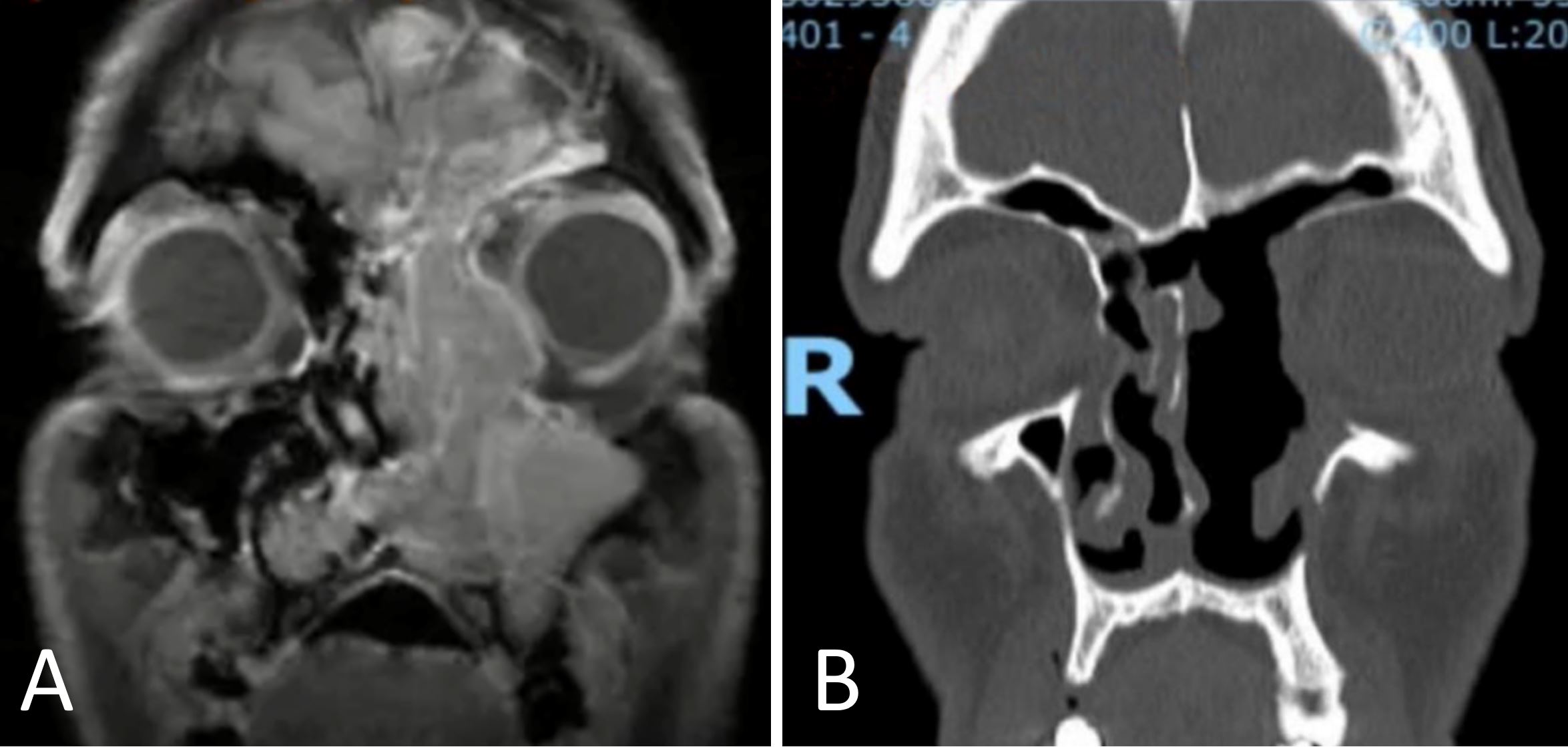
A 49-year-old male presented to the clinical office three months ago with left nasal obstruction, headaches, recurrent epistaxis, decreased visual acuity, and ipsilateral orbital cellulitis. A physical examination revealed that he had proptosis as well as a friable vegetative lesion that had obliterated the left nasal cavity. A magnetic resonance imaging of the paranasal sinuses (Figure 1A) revealed that a massive expansive lesion had occupied the left nasal cavity up to the level of the choanae, measuring 9.0 x 4.8 x 2.1 cm and displaying contrast enhancement. We decided not to perform an initial biopsy prior to surgery due to the high risk of bleeding and the intention to resect the lesion en bloc.


Figure 1. An imaging study displaying preoperative magnetic resonance imaging (A) and postoperative computed tomography (B).


The patient underwent an endoscopic procedure employing a centripetal technique. With this technique, the medial orbital wall and anterior base of the skull were preserved, while the lesion was completely removed. Following the operation, with open paranasal sinuses, there was no periorbital disease. However, a small leak of cerebrospinal fluid was noted within the ethmoidal fovea that was promptly closed using a free middle turbinate flap.

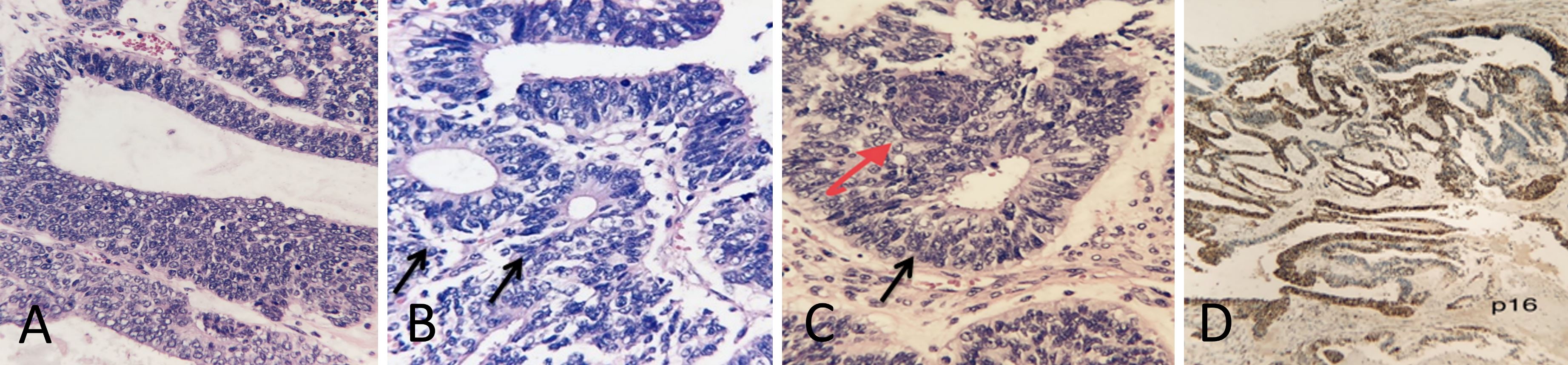
An immunohistochemical analysis (Figure 2) revealed a high-grade non-intestinal adenocarcinoma with 51 mitoses per 10 high-power field, positivity for CK7 and P16 antibodies and negativity for CK20, CDX2 and Ki-67 antibodies with a proliferative index of 70%. It was determined that the patient had a stage pT3N0M0 and was referred to the oncology and radiotherapy departments.


Figure 2. (A) In high-grade non-intestinal adenocarcinomas, glandular formation, marked cellular proliferation, and cellular atypia are evident (x10). (B) This is a high-grade non-intestinal adenocarcinoma characterized by glandular formation and some areas with Flexner-type rosettes (black arrows) and nuclear pleomorphism (x10). (C) The image shows a high-grade non-intestinal adenocarcinoma with a Morular-like metaplasia (red arrow), rosettes-like formation (black arrow), and nuclear atypia (x10). (D) A multi-focal and strong positivity is observed for p16 (x4).


Radiotherapy was administered as adjuvant therapy, but chemotherapy was not indicated. A total of 25 radiotherapy sessions were performed on him with a total dose of 50 Gy. It was decided to use intensity modulated radiotherapy because it is capable of administering higher and more effective doses to tumors with fewer side effects when compared to conventional radiotherapy. There were no significant side effects associated with radiotherapy. The patient underwent a periodic follow-up procedure with serial computed tomography scans on a quarterly basis. There was no evidence of recurrence of the disease after 30 months of treatment, and he continues to follow up with an outpatient physician on a regular basis.

Discussion

Only a few reports of high-grade sinonasal adenocarcinomas of the non-intestinal type have been published [1,10]. Males are more susceptible to the disease, and it can impact individuals of all ages, from adolescents to the elderly [1,4,10]. As shown in this case, nasal obstruction is the most common symptom, which may be accompanied by swelling or facial deformities. The most common sites of involvement are the nasal cavity and maxillary sinuses [1,7,10,11], although there are instances, such as this one, in which it can extend to other sinuses as well. Adenocarcinomas of the intestinal type have been associated with occupational exposures to wood dust, leather, and flour, whereas there are no risk factors identified for high-grade non-intestinal adenocarcinomas [10].

Even with aggressive treatment, high-grade tumors have a poor prognosis, with a survival rate of only 20% after three years [4,9]. Historically, the average 5-year survival rates of sinonasal carcinomas have been reported to be 28% in the 1960s and 51% in the 1990s, respectively [3]. A recent study by Turner et al. showed that there were some incremental improvements that approached statistical significance [3]. Based on these findings, it has been estimated that the 5-year relative survival rate has increased from approximately 49.7% for patients diagnosed in 1973 to 56.4% for patients diagnosed in 2001. Based on the analysis of 418 patients by Choussy et al., a 5-year overall survival rate of 64% was reported [12].

In a study performed by Bhayani et al., 66 patients were examined, 31 of whom had non-intestinal adenocarcinoma, and a 5-year overall survival rate of 65.9% was reported [13]. Among 24 patients studied by Orvidas et al., 58% had non-intestinal adenocarcinomas, and the 5-year overall survival rate was 58% [14]. The study conducted by Chen et al. is the only one to independently analyze non-intestinal adenocarcinoma [15]. In an analysis of 300 cases of non-intestinal adenocarcinoma, the authors reported a disease-specific survival rate of 71.2% with no statistically significant differences in survival when compared with 25 cases of intestinal adenocarcinoma. In a study of low-grade and high-grade sinonasal non-intestinal-type adenocarcinomas, the 5-year overall survival rate and the disease-specific survival rate were 100% and 100% for G1 tumors, respectively; 87.5% and 87.5% for G3 tumors, respectively; while the 5-year recurrency-free survival rate was 91.7% for G1 tumors and 88.9% for G3 tumors [9].

As far as the nasal cavity and the paranasal sinuses are concerned, adenocarcinomas can be classified as salivary gland, intestinal, or non-intestinal types of low or high grade [10,16]. The non-intestinal type uses these nomenclatures due to the lack of morphological characteristics and immunohistochemical patterns observed in salivary gland adenocarcinomas or intestinal type adenocarcinomas [4,17,18].

Upon macroscopy, non-intestinal tumors may have flat or exophytic appearances, and their color may range from white to violaceous, and they may contain areas that are friable and/or solid, depending on the type of tumor [10,18]. In the case described, macroscopically, there was an irregular outline associated with exophytic growth, violaceous color, and areas that were sometimes friable and sometimes solid.

Low-grade non-intestinal adenocarcinoma is an encapsulated tumor with a pattern of glandular or papillary growth. It presents low to moderate pleomorphism in the cell structure, with occasional mitoses, but not with atypical mitoses or necrosis [1,17,18].

In contrast, high-grade non-intestinal adenocarcinoma is an invasive tumor that has a solid pattern, containing glandular or papillary foci; some of the tumors exhibit blastomatous characteristics of teratocarcinoma [10,17], may form Flexner rosettes, and may exhibit morular squamous metaplasia, as shown in Figures 2A and 2B, respectively in this report. There is a moderate to severe level of pleomorphism, and the mitotic index is high (more than 5 mitotic figures per 10 high-power fields), characterized by both atypical mitosis and necrosis [4,10,18]. It is imperative to distinguish between salivary gland-type adenocarcinoma, intestinal-type adenocarcinoma, and metastatic adenocarcinoma when making the differential diagnosis [18].

An immunohistochemical pattern of high-grade non-intestinal adenocarcinoma shows a strong and diffuse positivity for CK7 [16,17]. There may be some cases where p16 is positive (as shown in Figure 2D in our case report), which necessitates the detection of HPV strains with a high likelihood of causing neoplasia. Rarely, neuroendocrine markers such as synaptophysin and chromogranin exhibit weak or focal positivity [17]. They are negative for CK20 and CDX-2, which differentiates them from intestinal type adenocarcinoma and defines the final diagnosis in this case [16,18].

When adenocarcinomas are detected at an early stage, surgical treatment can be used alone, resulting in a 5-year survival rate of 83.4%. Among patients with more advanced stages of the disease, the 5-year survival rate is 66.6% when surgery and radiotherapy are combined. Radiotherapy alone was not found to have a significant benefit in terms of 5-year survival when compared with no treatment [19].

There are some key insights that can be gained from this case. To begin with, high-grade, non-intestinal sinonasal adenocarcinomas are uncommon and aggressive. It is common for patients to experience nasal obstruction as one of the most prominent symptoms. A complete resection of the tumor may result in better outcomes, which can be achieved through endoscopic access. A nonspecific clinical presentation usually results in a delayed diagnosis. A positive immunohistochemistry finding was observed for CK 7 and a negative finding for CK20 and CDX-2. In this case, we learned that high-grade non-intestinal adenocarcinoma is an exclusion diagnosis, because the histopathological characteristics are used to identify and exclude other forms of adenocarcinoma, including those of the intestinal and salivary types. A multidisciplinary assessment is also essential for the effective management of patients. The purpose of this case report is to emphasize that surgery with meticulous resection is essential to ensure a better outcome.

Conclusion

This is an uncommon case of high-grade non-intestinal sinonasal adenocarcinoma that represents a challenging diagnosis. It is imperative to distinguish it from other sinonasal tumors since it has a significantly different prognosis. For a definitive diagnosis, a detailed anamnesis is required, a radical endoscopic resection to achieve a total excision is necessary, and satisfactory immunohistochemistry results are required.

References

  1. Pradhan P, Panigrahi R, Misra P, Senapati U, Samantaray K. High-grade non-salivary non-intestinal adenocarcinoma of the nasal cavity - A less known entity. Int J Otorhinolaryngol Head Neck Surg 2020;6(11):2163-2164. [View Article]
  2. Flint PW, Haughey BH, Robbins KT, et al. Cummings Otolaryngology: Head and Neck Surgery. 6th ed. Philadelphia: Elsevier Saunders;2015.
  3. Turner JH, Reh DD. Incidence and survival in patients with sinonasal cancer: A historical analysis of population-based data. Head Neck 2012;34(6):877-885. [View Article]
  4. Heffner DK, Hyams VJ, Hauck KW, Lingeman C. Low-grade adenocarcinoma of the nasal cavity and paranasal sinuses. Cancer 1982;50(2):312-322. [View Article]
  5. Breda M, Miranda D, Pereira S, et al. [Sinonasal adenocarcinoma - Hospital de Braga ENT department expertise]. Port J Otorhinolaryngol Head Neck Surg 2017;54(4):233-238. [View Article]
  6. Vergez S, du Mayne MD, Coste A, et al. Multicenter study to assess endoscopic resection of 159 sinonasal adenocarcinomas. Ann Surg Oncol 2014;21(4):1384-1390. [View Article]
  7. Lund VJ, Stammberger H, Nicolai P, et al. European position paper on endoscopic management of tumours of the nose, paranasal sinuses and skull base. Rhinol Suppl 2010;22:1-143. [View Article]
  8. Madani I, Bonte K, Vakaet L, Boterberg T, De Neve W. Intensity-modulated radiotherapy for sinonasal tumors: Ghent University Hospital update. Int J Radiat Oncol Biol Phys 2009;73(2):424-432. [View Article]
  9. Bignami M, Lepera D, Volpi L, et al. Sinonasal Non-intestinal-type adenocarcinoma: A retrospective review of 22 patients. World Neurosurg 2018;120:e962-e969. [View Article]
  10. Stelow EB, Mills SE, Jo VY, Carlson DL. Adenocarcinoma of the upper aerodigestive tract. Adv Anat Pathol 2010;17(4):262-269. [View Article]
  11. Jain C, Caulley L, Macdonald KI, et al. Nasopharyngeal non-intestinal-type adenocarcinoma: A case report and updated review of the literature. Curr Oncol 2017;24(1):e55-e60. [View Article]
  12. Choussy O, Ferron C, Vedrine PO, et al. Adenocarcinoma of ethmoid: A GETTEC retrospective multicenter study of 418 cases. Laryngoscope 2008;118(3):437-443. [View Article]
  13. Bhayani MK, Yilmaz T, Sweeney A, et al. Sinonasal adenocarcinoma: A 16-year experience at a single institution. Head Neck 2014;36(10):1490-1496. [View Article]
  14. Orvidas LJ, Lewis JE, Weaver AL, Bagniewski SM, Olsen KD. Adenocarcinoma of the nose and paranasal sinuses: A retrospective study of diagnosis, histologic characteristics, and outcomes in 24 patients. Head Neck 2005;27(5):370-375. [View Article]
  15. Chen MM, Roman SA, Sosa JA, Judson BL. Predictors of survival in sinonasal adenocarcinoma. J Neurol Surg B Skull Base 2015;76(3):208-213. [View Article]
  16. Cathro HP, Mills SE. Immunophenotypic differences between intestinal-type and low-grade papillary sinonasal adenocarcinomas: An immunohistochemical study of 22 cases utilizing CDX2 and MUC2. Am J Surg Pathol 2004;28(8):1026-1032. [View Article]
  17. Mills SE, Greenson JK, Hornick JL, Longacre TA, Reuter VE. Sternberg's Diagnostic Surgical Pathology. 6th ed. Philadelphia: Wolters Kluwer;2015.
  18. Fletcher CDM. Diagnostic Histopathology of Tumors. 5th ed. Philadelphia: Elsevier;2020.
  19. Kilic S, Samarrai R, Kilic SS, Mikhael M, Baredes S, Eloy JA. Incidence and survival of sinonasal adenocarcinoma by site and histologic subtype. Acta Otolaryngol 2018;138(4):415-421. [View Article]

Editorial Information

Publication History

Received date: May 01, 2022
Accepted date: August 18, 2022
Published date: September 22, 2022

Disclosure

The manuscript has not been presented at any meetings on the topic.

Ethics Approval and Consent to Participate

The study is in accordance with the ethical standards of the 1964 Helsinki declaration and its later amendments or comparable ethical standards. The authors obtained permission from the participants in the human research prior to publishing their images or photographs.

Funding

This research has received no specific grant from any funding agency either in the public, commercial, or not-for-profit sectors.

Conflict of Interest

There are no conflicts of interest declared by either the authors or the contributors of this article, which is their intellectual property.

Publisher Disclaimer

It should be noted that the opinions and statements expressed in this article are those of the respective author(s) and are not to be regarded as factual statements. These opinions and statements may not represent the views of their affiliated organizations, the publishing house, the editors, or any other reviewers since these are the sole opinion and statement of the author(s). The publisher does not guarantee or endorse any of the statements that are made by the manufacturer of any product discussed in this article, or any statements that are made by the author(s) in relation to the mentioned product.

Copyright

© 2022 The Author(s). This is an open-access article distributed under the terms of the Creative Commons Attribution 4.0 International License (CC-BY). In accordance with accepted academic practice, anyone may use, distribute, or reproduce this material, so long as the original author(s), the copyright holder(s), and the original publication of this journal are credited, and this publication is cited as the original. To the extent permitted by these terms and conditions of license, this material may not be compiled, distributed, or reproduced in any manner that is inconsistent with those terms and conditions.

Department of Otolaryngology-Head and Neck Surgery, Santa Casa de Misericordia Hospital, Federal University of Health Sciences of Porto Alegre, Porto Alegre, Rio Grande do Sul, Brazil
Department of Pathology, Hospital de Clínicas de Porto Alegre, Federal University of Rio Grande do Sul, Porto Alegre, Rio Grande do Sul, Brazil
Department of Otolaryngology-Head and Neck Surgery, Santa Casa de Misericordia Hospital, Federal University of Health Sciences of Porto Alegre, Porto Alegre, Rio Grande do Sul, Brazil
Email: b_beraldin@hotmail.com
Address: Rua Professor Annes Dias, 295. Centro Histórico, Porto Alegre, RS 90020-090, Brazil
Figure 1.JPG
Figure 1. An imaging study displaying preoperative magnetic resonance imaging (A) and postoperative computed tomography (B).
Figure 2.JPG
Figure 2. (A) In high-grade non-intestinal adenocarcinomas, glandular formation, marked cellular proliferation, and cellular atypia are evident (x10). (B) This is a high-grade non-intestinal adenocarcinoma characterized by glandular formation and some areas with Flexner-type rosettes (black arrows) and nuclear pleomorphism (x10). (C) The image shows a high-grade non-intestinal adenocarcinoma with a Morular-like metaplasia (red arrow), rosettes-like formation (black arrow), and nuclear atypia (x10). (D) A multi-focal and strong positivity is observed for p16 (x4).

Reviewer 1 Comments

  1. An unusual case of high-grade non-intestinal sinonasal adenocarcinoma is presented in this report. This case is of particular interest because it represents an aggressive and rare disease treated with excellent results. It is concluded that it is imperative to distinguish this tumor from other sinonasal tumors, since it has a significantly different prognosis. The authors propose that a detailed anamnesis, endoscopic resection to achieve complete excision, and satisfactory immunohistochemical analysis are required to confirm the diagnosis. Generally, I consider the case report to be informative, however, some details need to be clarified.
    ResponseThank you for these valuable comments.
     
  2. There is a clear consensus that the recommended treatment for adenocarcinomas is surgery followed by postoperative radiotherapy. The use of postoperative radiotherapy for sinonasal adenocarcinomas is not as clear as it should be. However, the local control rates of combined treatment strategies for advanced cases are comparable to those achieved in less advanced cases with surgery alone. This suggests that radiotherapy may have some value in this situation. In the case of tumors that are highly staged (stages T3 and T4) and of high grade, adjuvant radiotherapy can prove to be beneficial. In the present case, the details of the patient's adjuvant radiotherapy and/or chemotherapy are unclear. A more detailed description of the postoperative treatment regimen is required.
    ResponseThank you for your valuable comment. Included in the case presentation: He received adjuvant radiotherapy but chemotherapy was not indicated. He was submitted to 25 radiotherapy applications, with a total 50Gy dose. Intensity modulated radiotherapy was chosen due to the possibility of administering larger and more effective doses to the tumor with fewer side effects, compared to conventional radiotherapy techniques. No significant side effects from radiotherapy were observed.
     
  3. Prior to designing a cancer treatment plan, it is important to have information on TNM stages. What stage of cancer did this patient possess according to the American Joint Committee on Cancer (AJCC) TNM system?
    ResponseThank you for your valuable comment. Included in the case presentation: The patient was classified as stage pT3N0M0 and was referred to oncology and radiotherapy.
     
  4. The Abstract should include a short discussion of the lessons that might be brought forth by the case report. This type of discussion could be of significant assistance to researchers in assessing the potential interest of a particular case report.
    ResponseThank you for your valuable comment. We have included this information in the 14th paragraph of the discussion: With this case, we learned that high-grade non-intestinal adenocarcinoma is a diagnosis of exclusion since we need the histopathological characteristics to define and exclude other forms of adenocarcinomas such as intestinal and salivary types.
     
  5. It is noteworthy that this case report describes a rare occurrence. Nevertheless, the rarity of the case itself may not justify publication. In order to gain a deeper understanding of the unique nature of this case and how it contributes to the existing literature, the authors should discuss any potentially required modifications to clinical practice or diagnostic/prognostic methods.
    ResponseThank you for your valuable comment. In the last paragraph of the introduction, we justify the publication of the article: The report of this case is justified by the fact that it presents a rare and aggressive disease treated with excellent evolution. This suggests that a well performed surgery, with complete removal of the lesion, as performed in this case, seems to result in a better prognosis. In this way, it can contribute to understanding and treatment, benefiting other patients. Cases like this are little discussed in the literature and have several knowledge gaps.
     
  6. The Discussion indicates that despite aggressive treatment, more than 50% of patients die. There is no clear indication as to whether this means 50% of patients may die after surgery or a period of treatment after surgery. The issue needs to be clarified in a more concrete manner, for example, through a 5-year survival rate.
    ResponseThank you for your valuable comment. Survival rate information had not really been clearly included. We have included prognostic information, but the specific prognostic information in the high-grade non-intestinal type in the literature is unclear. We included in the discussion: Despite aggressive treatment, high-grade tumors have a poor prognosis, with an estimated 3-year survival rate of 20%. Historically, average overall 5-year survival rates of sinonasal carcinomas were reported at 28% in the 1960s and 51% in the 1990s. Turner et al. in a recent data showed some incremental improvement that closely approaches statistical significance. This corresponds to a change in 5-year relative survival from approximately 49.7% for patients diagnosed in 1973 to 56.4% for those diagnosed in 2001. Choussy et al. considered 418 patients and reported a 5-year overall survival rate of 64%. Bhayani et al. considered 66 patients, 31 of whom had non-intestinal adenocarcinoma, and reported a 5-year overall survival rate of 65.9%. Orvidas et al. considered 24 patients, of whom 58% had non-intestinal adenocarcinoma, and reported a 5-year overall survival of 58%. The study by Chen et al. is the only one that analyzed non-intestinal adenocarcinoma independently. These authors reported a disease specific survival rate of 71.2% in 300 cases of non-intestinal adenocarcinoma with no differences in terms of survival compared with 25 cases of intestinal adenocarcinoma. Low-grade and high-grade tumors were compared, and 5-year overall survival and disease specific survival were 100% for G1 and 87.5% for G3, and 5-year recurrency-free survival was 91.7% for G1 and 88.9% for G3.

Reviewer 2 Comments

  1. The present report documents an unusual case of high-grade non-intestinal sinonasal adenocarcinoma. This case involves a rare and aggressive disease that was successfully treated. According to the authors, it is imperative to distinguish this tumor from other sinonasal tumors since it is associated with a significantly different prognosis. For a diagnosis to be confirmed, the authors recommend a thorough history, endoscopic resection for complete excision, and satisfactory immunohistochemical analysis. A thorough reading of the case report is recommended, although some points need clarification.
    ResponseThank you for these valuable comments.
     
  2. In the first paragraph of the Introduction, there is some confusion. As it is mentioned, non-salivary nasosinusal adenocarcinomas comprise 13% of carcinomas. Does the term "carcinoma" encompass all cancers, or only those arising in the sinonasal region? My suggestion is that the authors should rephrase it in a more understandable manner as follows: Sinonasal adenocarcinomas can be divided into salivary-type and non-salivary-type adenocarcinomas. The latter are further divided into intestinal-type and nonintestinal-type adenocarcinomas. Sinonasal adenocarcinomas of the non-salivary type are rare cancers. They represent only 13% of all sinonasal carcinomas reported [Turner JH, Reh DD. Incidence and survival in patients with sinonasal cancer: a historical analysis of population-based data. Head Neck 2012;34(6):877-885].
    ResponseThank you for your valuable comment. Suggestion accepted, we added bibliography and changed to: The sinonasal region is commonly related to a series of tumors, but the occurrence of adenocarcinomas is uncommon. Sinonasal adenocarcinomas can be divided into salivary-type and non-salivary-type adenocarcinomas. The latter are further divided into intestinal-type and nonintestinal-type adenocarcinomas. Sinonasal adenocarcinomas of the non-salivary type are rare cancers. They represent only 13% of all sinonasal carcinomas reported.
     
  3. It is somewhat unclear in the description of the surgical procedure that began with, "The procedure was performed using a centripetal technique, respecting limits, the medial wall of the orbit and the anterior skull base, with complete resection of the lesion". What type of surgery did you undergo? Was it an endoscopic resection? For a better understanding of the surgical procedure, please provide more detail.
    ResponseThank you for your valuable comment. We added this information: The patient was submited to an endoscopic procedure, which was performed using a centripetal technique, respecting the limits, the medial wall of the orbit and the anterior base of the skull, with complete resection of the lesion.
     
  4. The authors should provide more detailed information regarding oncological treatments, including adjuvant radiotherapy and chemotherapy. Was there any adverse reaction to the treatment? Moreover, the follow-up period from the date of surgery to the present and the final pathologic TNM stage should also be provided.
    ResponseThank you for your valuable comment. We added this information: The patient was classified staging pT3N0M0 and was referred to oncology and radiotherapy. He received adjuvant radiotherapy but chemotherapy was not indicated. He was submited to 25 radiotherapy applications, with a total 50Gy dose. Intensity modulated radiotherapy was chosen due to the possibility of administering larger and more effective doses to tumors with fewer side effects compared to conventional radiotherapy techniques. No significant side effects from radiotherapy were observed. He was submitted to quarterly follow-up with serial computed tomography. After 30 months, he remains free of disease recurrence and follows outpatient follow-up.
     
  5. It is advised that the key points of the case report should be summarized in the Discussion section. Particularly, it may be useful for readers to know how a similar situation might be handled differently in the case report. Having a summary of the main points will assist researchers in extracting the most value from the article's contente.
    ResponseThank you for your valuable comment. We added this information at the end of the discussion: High-grade, non-intestinal type sinonasal adenocarcinomas are rare and aggressive tumors. The most prevalent symptom is nasal obstruction. Diagnosis is usually delayed due to nonspecific clinical presentation. Complete ressection of the tumor may lead to better outcomes, and can be achieved through endoscopic access. Immunohistochemistry is positive for CK7 and negative for CK20 and CDX-2.
     
  6. As stated in the Discussion, "the latest two classifications” use these nomenclatures because they do not have the morphological characteristics and immunohistochemical pattern of salivary gland adenocarcinoma or intestinal type adenocarcinoma. It would be very helpful if you could clarify what "the latest two classifications" mean.
    ResponseThank you for your valuable comment. We made changes to make it more clear: The non-intestinal type uses these nomenclatures because they do not have morphological characteristics and immunohistochemical pattern of salivary gland adenocarcinoma or intestinal type adenocarcinoma.
     
  7. The conclusion emphasizes the importance of distinguishing this tumor from other sinonasal tumors, which have a markedly different prognosis. Nonetheless, the prognosis of this disease was not adequately addressed, and a detailed discussion is required, in particular how this disease differs from other sinonasal tumors. The authors are encouraged to also discuss overall survival rates and disease-free survival rates as prognosis issues. Furthermore, does surgery followed by radiotherapy or chemotherapy enhance the likelihood of a disease-free outcome compared to surgery alone?
    ResponseThank you for your valuable comment. Survival rate information had not really been clearly included. We have included prognostic information, but the specific prognostic information in the high-grade non-intestinal type in the literature is unclear. About the treatments, there is no clear evidence in literature whether associating chemotherapy or radiotherapy brings better outcomes. We included in the discussion: Despite aggressive treatment, high-grade tumors have a poor prognosis, with an estimated 3-year survival rate of 20%. Historically, average overall 5-year survival rates of sinonasal carcinomas were reported at 28% in the 1960s and 51% in the 1990s. Turner et al. in a recent data showed some incremental improvement that closely approaches statistical significance. This corresponds to a change in 5-year relative survival from approximately 49.7% for patients diagnosed in 1973 to 56.4% for those diagnosed in 2001. Choussy et al. considered 418 patients and reported a 5-year overall survival rate of 64%. Bhayani et al. considered 66 patients, 31 of whom had non-intestinal adenocarcinoma, and reported a 5-year overall survival rate of 65.9%. Orvidas et al. considered 24 patients, of whom 58% had non-intestinal adenocarcinoma, and reported a 5-year overall survival of 58%. The study by Chen et al. is the only one that analyzed non-intestinal adenocarcinoma independently. These authors reported a disease specific survival rate of 71.2% in 300 cases of non-intestinal adenocarcinoma with no differences in terms of survival compared with 25 cases of intestinal adenocarcinoma. Low-grade and high-grade tumors were compared, and 5-year overall survival and disease specific survival were 100% for G1 and 87.5% for G3, and 5-year recurrency-free survival was 91.7% for G1 and 88.9% for G3.

Reviewer 3 Comments

  1. The present case report describes the successful treatment of an unusually aggressive form of non-intestinal sinonasal adenocarcinoma in a 49-year-old male patient. It is argued that the differentiation of this tumor from other sinonasal tumors is of paramount importance, due to its significantly different prognosis. The authors recommend a comprehensive history, an endoscopic resection to guarantee complete removal, and a satisfactory immunohistochemical analysis to confirm the diagnosis. In my opinion, there are a few issues that need to be addressed before this article can be considered for publication.
    ResponseThank you for these valuable comments.
     
  2. It has been shown that sinonasal adenocarcinomas are commonly associated with occupational or environmental carcinogens, such as wood dust and Epstein-Barr virus. According to the International Agency for Research on Cancer (IARC), a number of physical-chemical agents have been classified as having carcinogenic effects on sinonasal cancer. There is a very low probability of developing sinonasal adenocarcinoma in an individual without risk factors. In the current case, there is a lack of clarity in the description of the current medical condition and the medical history. There is a need to describe whether the patient was exposed to occupational carcinogens, tobacco smoke and alcohol.
    ResponseThank you for your valuable comment. Information has been included: Intestinal type adenocarcinomas have association with occupational exposure, like wood dust, leather and flour, while there were no identified risk factors for high-grade non-intestinal adenocarcinomas.
     
  3. It should be detailed as to the clinical examinations that were performed. For instance, was a biopsy performed prior to the surgery?
    ResponseThank you for your valuable comment. Information has been included: It was chosen not to perform a biopsy prior to surgery due the high risk of bleeding and the intention to resect the lesion en bloc during surgery.
     
  4. In order to be effective in eradicating the disease, it is essential to select the surgical approach that allows for complete excision of the tumor. There is some evidence that endoscopic techniques, if possible, are comparable to external techniques, if not superior, in terms of complete removal of tumors. In this case, was the endoscopic approach considered the surgery of choice after it was appropriately planned and justified?
    ResponseThank you for your valuable comment. Information has been included: There are a variety of external approach possibilities, but endoscopic surgery has gained special prominence in many centers, since it offers similar oncological results, lower rate of complications and lower mortality associated with surgical procedure. The patient was submited to an endoscopic procedure, which was performed using a centripetal technique, respecting the limits, the medial wall of the orbit and the anterior base of the skull, with complete resection of the lesion.
     
  5. It would be useful for the authors if they could compare the case report to the literature. When the current findings do not accord with the published literature, possible explanations should be offered.
    ResponseThank you for your valuable comment. Information has been included: The most prevalent symptom is nasal obstruction, which may be associated with swelling or facial deformity, as presented on this case. In the case described, macroscopically, the lesion had irregular outline with exophytic growth, violaceous color and areas that were sometimes friable and sometimes solid. High-grade non-intestinal adenocarcinoma, on the other hand, is an invasive tumor with a solid pattern, which may contain focus of a glandular or papillary pattern; some have areas that resemble the blastomatous component of a teratocarcinoma, may form Flexner type rosettes and areas with morular squamous metaplasia, as illustrated in Figures 2B and 2C in the description of the report. The immunohistochemical pattern of high-grade non-intestinal adenocarcinoma shows a strong and diffuse positivity for CK7, some cases may have a positive p16 (shown with Figure 2D in our case report).
     
  6. It is suggested that there is a section in the Discussion that discusses the experiences that can be learned from the case report. A section on this would be a valuable addition to the medical knowledge as well as educational content.
    ResponseThank you for your valuable comment. Information has been included: With this case, we learned that high-grade non-intestinal adenocarcinoma is a diagnosis of exclusion since we need the histopathological characteristics to define and exclude other forms of adenocarcinomas such as intestinal and salivary types. In addition, a multidisciplinary assessment is essential for patient management. This case report reiterates that surgery with meticulous resection is essential for a better evolution.
     
  7. There is a mention in the Discussion that “some have cystic areas”. The statement is vague and needs clarification.
    ResponseThank you for your valuable comment. We have decided to remove this information.

Editorial Comments

  1. There is a requirement that the names of institutions of the authors have to be translated into English.
    ResponseThank you for your valuable comment. We transleted it into English.
     
  2. It is appropriate to indicate the credentials of the authors, such as MD, PhD.
    ResponseThank you for your valuable comment. We have included this information.
     
  3. The term "nasosinusual" adenocarcinoma should be replaced with "sinonasal" adenocarcinoma in the text.
    ResponseThank you for your valuable comment. We have replaced "nasosinusual" throughout the text with "sinonasal".
     
  4. There should be a sequential numbering of the figures in the text.
    ResponseThank you for your valuable comment. We added it.
     
  5. A clear indication of the magnification of the pathological images should be provided (Figures 3-6).
    ResponseThank you for your valuable comment. We modified it.
     
  6. Reference 4 needs to be translated into English so that it can be used for research and academic purposes.
    ResponseThank you for your valuable comment. We transleted the reference 4 into English.
     
  7. Reference 12 is not cited in the text of this paper.
    ResponseThank you for your valuable comment. We removed this reference, because it wasn’t really cited in the text.
     
  8. Reference 13 should also be placed within the text in numerical order, namely immediately following Reference 12.
    ResponseThank you for your valuable comment. We modified it so that the references are numbered sequentially.

Gehrke V, Beraldin BS, Lubianca Neto JF, Barra MB, Viga JDC, Vieira MZDD, Houdali ISHM, Rocha Filho MAM. A rare case of high-grade non-intestinal sinonasal adenocarcinoma. Arch Otorhinolaryngol Head Neck Surg. 2022;6(2):2. https://doi.org/10.24983/scitemed.aohns.2022.00165